Ear & Temporal Bone Tumor Surgery — Expert Care at Medanta Noida
Dr. Vimmi Gautam is a Head & neck cancer surgeon at Medanta Noida with specialized expertise in ear and temporal bone tumor surgery.
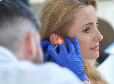
What Is Ear & Temporal Bone Tumor Surgery?
Ear and temporal bone tumor surgery refers to a spectrum of specialized surgical procedures designed to remove tumors that arise in or around the ear canal, middle ear, inner ear, and the temporal bone — the area of the skull that houses the hearing and balance organs, facial nerve, and major blood vessels.
These tumors may be benign (non-cancerous) or malignant (cancerous). Regardless of type, their location near the facial nerve (CN VII), jugular vein, and brain makes surgical precision critical. The goal of surgery is complete tumor removal while preserving hearing, facial movement, and quality of life wherever possible.
Types of Ear & Temporal Bone Tumors We Treat
We manage both benign and malignant ear and temporal bone tumors using advanced diagnostic techniques and specialized surgical approaches tailored to each patient.
Benign Tumors
Cholesteatoma
Abnormal skin growth in the middle ear that can damage surrounding structures.
Glomus Tumor
Highly vascular benign tumor arising from the middle ear or jugular bulb.
Acoustic Neuroma
Benign nerve sheath tumor affecting hearing and balance functions.
Osteoma
Benign bony growth developing within the external ear canal.
Inflammatory Polyps & Granulomas
Non-cancerous inflammatory growths that may cause ear symptoms and discomfort.
Malignant Tumors
Squamous Cell Carcinoma (SCC)
The most common cancer affecting the ear canal and temporal bone.
Basal Cell Carcinoma
Often begins on the outer ear and may invade deeper structures.
Adenoid Cystic Carcinoma
Slow-growing but locally aggressive cancer requiring specialized treatment.
Ceruminous Gland Adenocarcinoma
Rare malignant tumor arising from glands of the external ear canal.
Metastatic Tumors
Secondary spread of cancer from the breast, lung, prostate, kidney, or other organs.
Symptoms That May Indicate Tumors
Symptoms may vary based on location, but early recognition is crucial for timely diagnosis and treatment.
Lumps or Swelling
Persistent lumps or unusual swelling in the body
Pain or Discomfort
Ongoing pain in the affected area
Swallowing / Breathing Issues
Difficulty swallowing or breathing
Weight Loss
Unexplained or sudden weight loss
Voice Changes
Changes in voice or speech patterns
Non-Healing Ulcers
Sores or lesions that do not heal
Diagnosis & Treatment Planning
Before surgery, doctors perform detailed evaluations to determine the best treatment approach.
Clinical Examination
Initial physical assessment to evaluate symptoms and detect abnormalities.
Biopsy
Tissue sample is collected to confirm the type and nature of the tumor.
CT Scan / MRI
Imaging tests to determine tumor size, location, and spread.
PET Scan
Helps in staging the cancer and identifying spread to other parts of the body.
Blood Tests & Health Assessment
Evaluates overall health and fitness before planning surgery.
Recovery After Advanced Tumor Surgery
Recovery after advanced tumor removal requires careful monitoring, rehabilitation, and follow-up for optimal outcomes.
Hospital Stay
Typically 5–14 days depending on the complexity of surgery.
Gradual Recovery
Healing progresses gradually with continuous monitoring and medical care.
Rehabilitation
Physiotherapy or rehabilitation may be required for functional recovery.
Regular Follow-Up
Follow-ups help monitor healing and ensure cancer control.
Frequently Asked Questions
What is ear and temporal bone tumor surgery?
Ear and temporal bone tumor surgery is a specialised procedure used to remove benign or cancerous tumours from the ear canal, middle ear, inner ear, or temporal bone. The surgical approach depends on the tumour's size, location, and stage, ranging from a sleeve resection to a radical temporal bone resection.
What are the types of temporal bone resection surgery?
There are four main types: Sleeve Resection for outer ear canal tumors, Lateral Temporal Bone Resection for tumors involving the bony canal and middle ear, Subtotal Temporal Bone Resection for tumors behind the eardrum or inner ear, and Radical Temporal Bone Resection for advanced tumors involving the skull base or intracranial structures.
Will I lose my hearing after temporal bone tumor surgery?
Hearing outcomes depend on the type of surgery performed. Sleeve resection usually preserves hearing, while lateral temporal bone resection may require hearing aids afterward. Hearing loss on the operated side is generally expected after subtotal or radical temporal bone resection.
Is the facial nerve at risk during ear tumor surgery?
Yes, the facial nerve travels through the temporal bone and may be affected by surgery. Advanced microsurgical techniques and intraoperative nerve monitoring (IONM) help identify and protect the nerve. In selected cases, facial nerve reanimation procedures may be performed if nerve sacrifice is necessary.
How is a temporal bone tumour diagnosed?
Diagnosis typically includes clinical examination, otoscopy, high-resolution CT (HRCT), contrast-enhanced MRI, PET-CT for cancer staging, biopsy for tissue confirmation, and an audiogram to assess hearing before treatment planning.